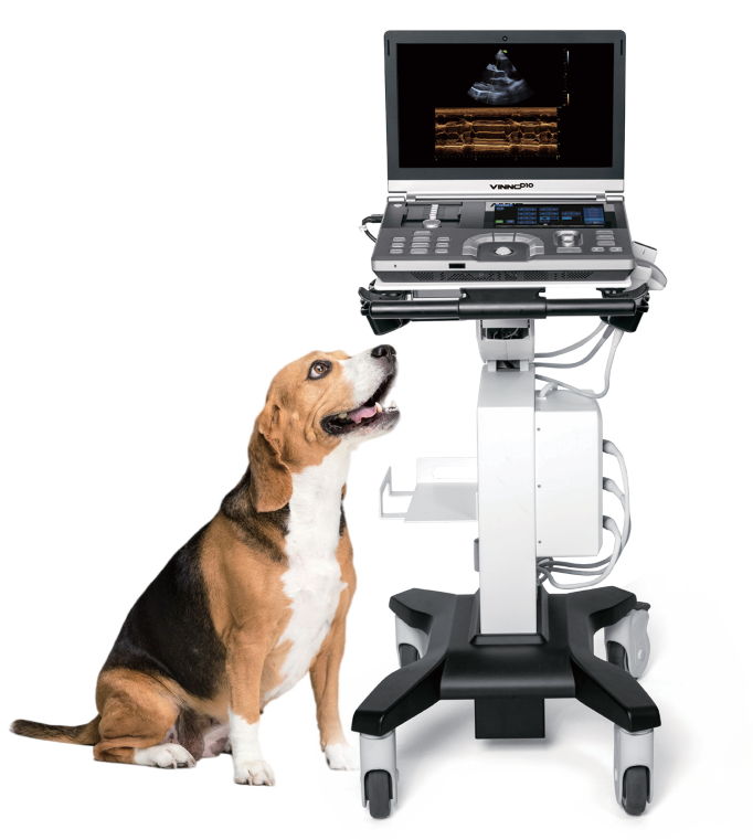
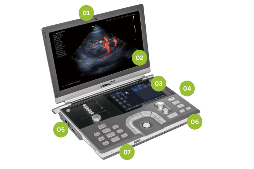
VINNO D10
Premium Compact Ultrasound Wherever You Need It
Advanced Beam Correction Technology

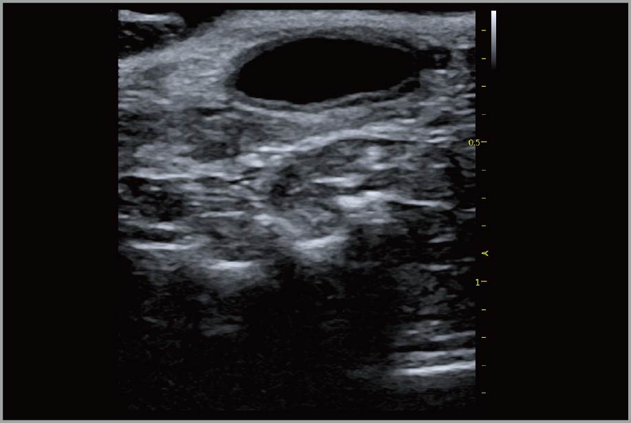
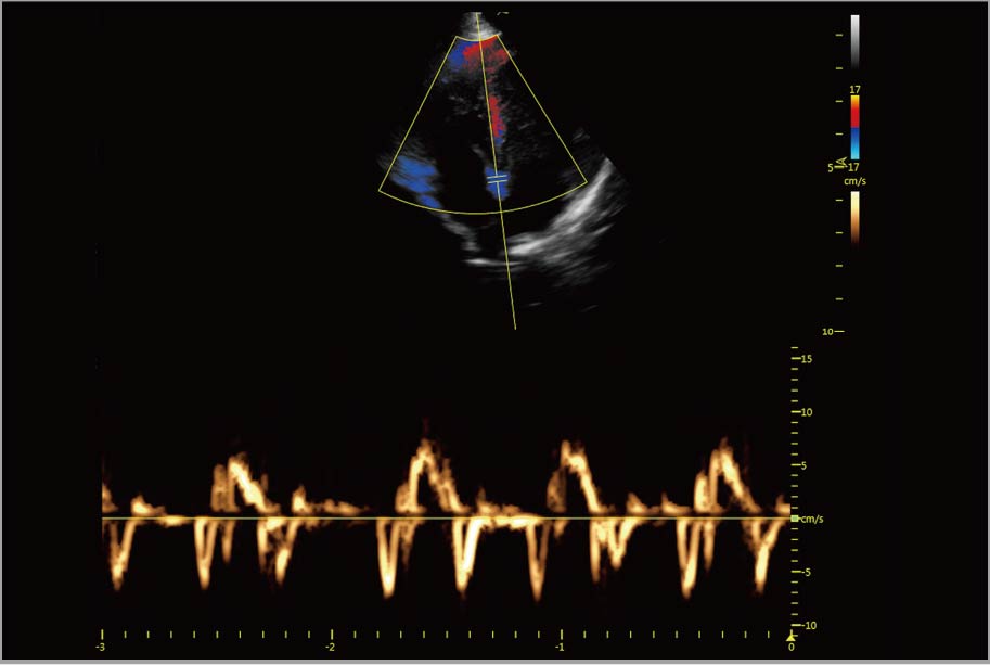
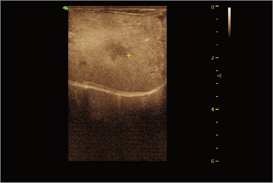
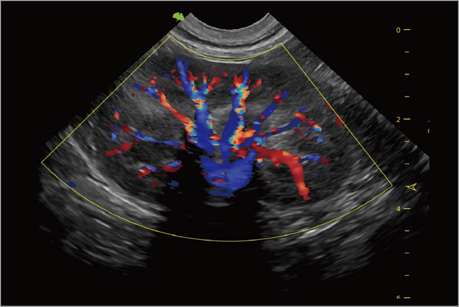
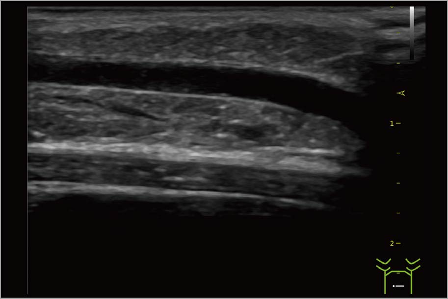
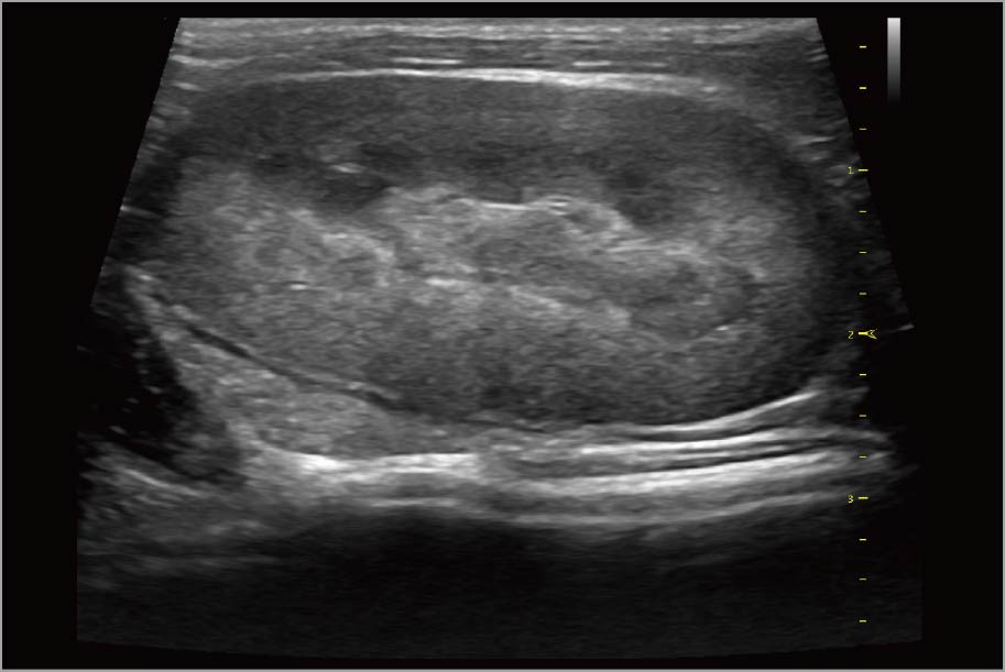
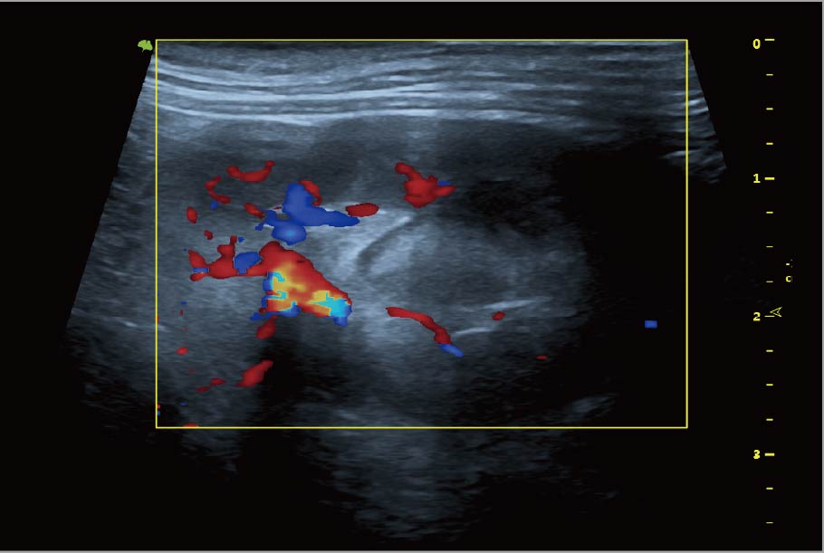
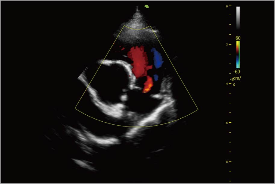
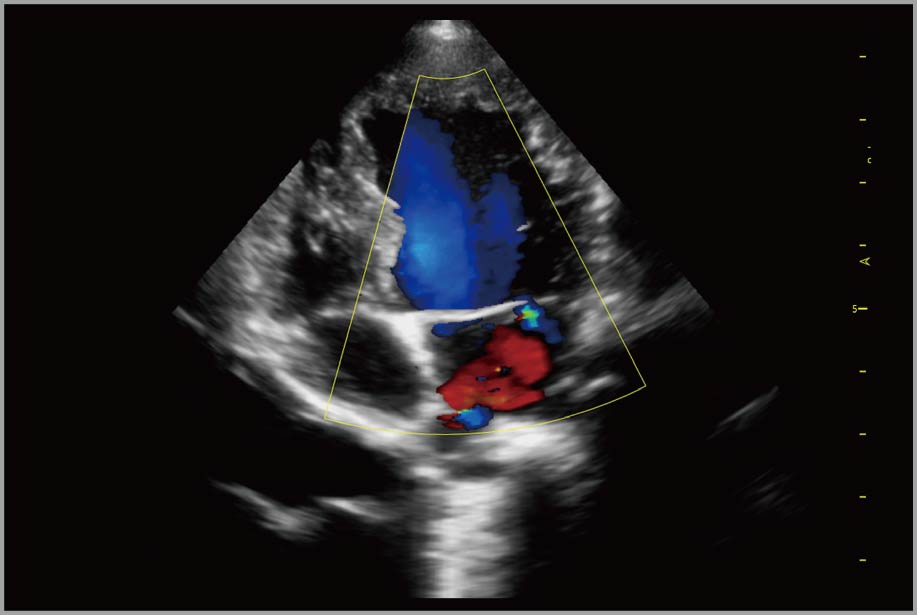
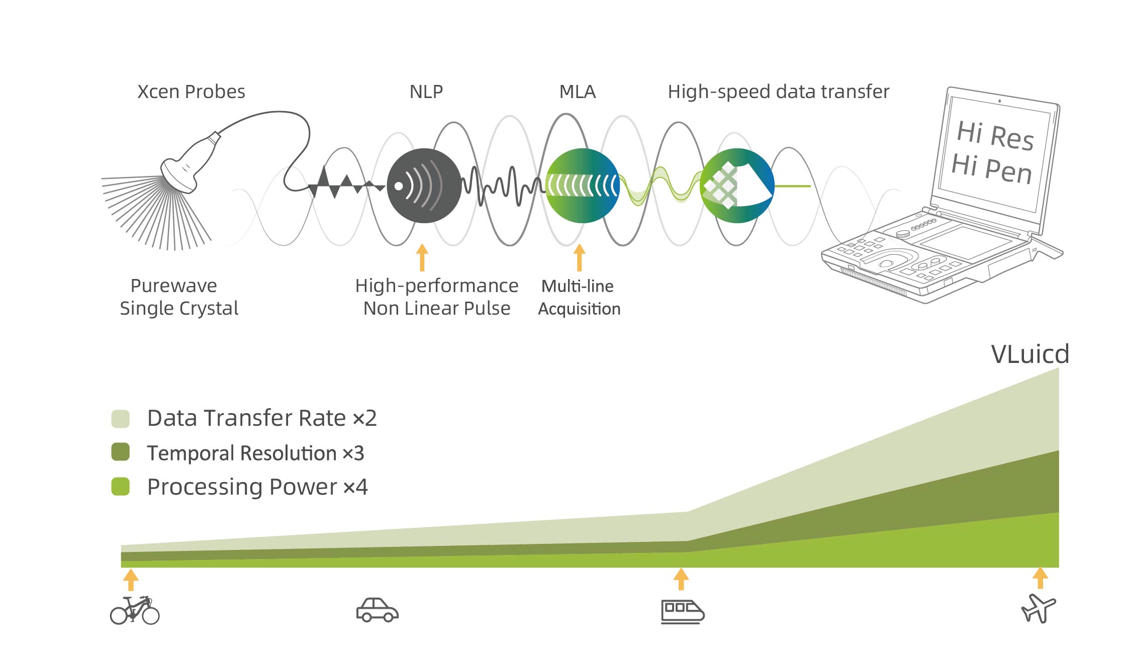
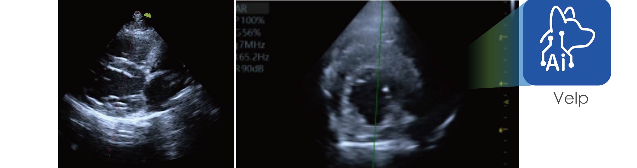
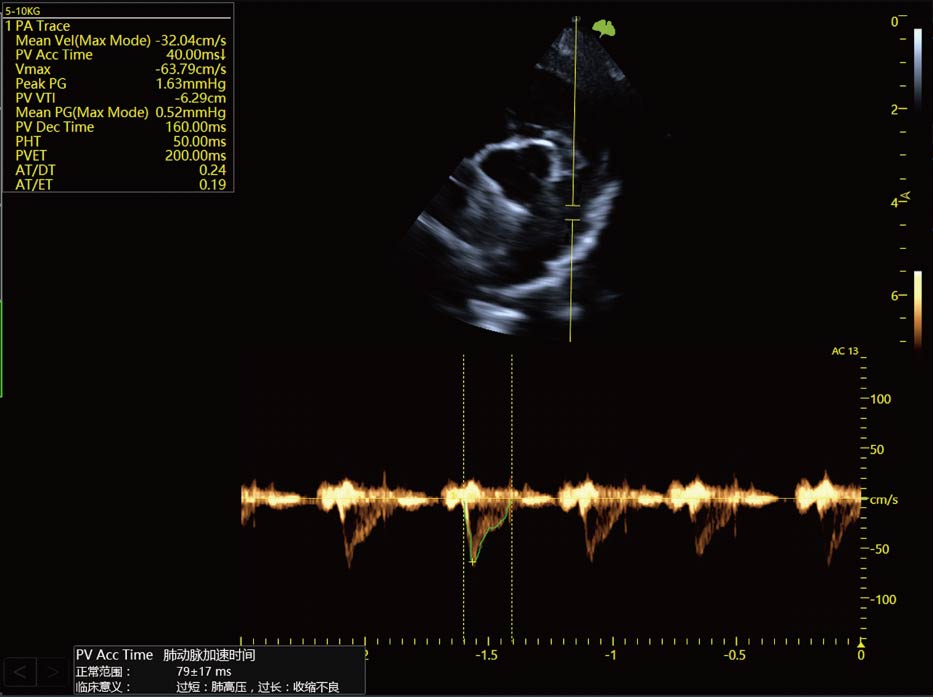
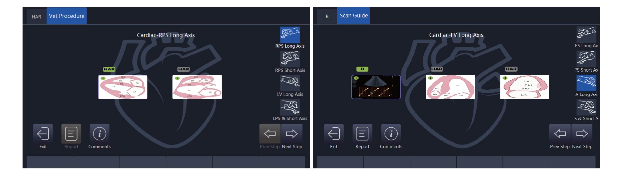
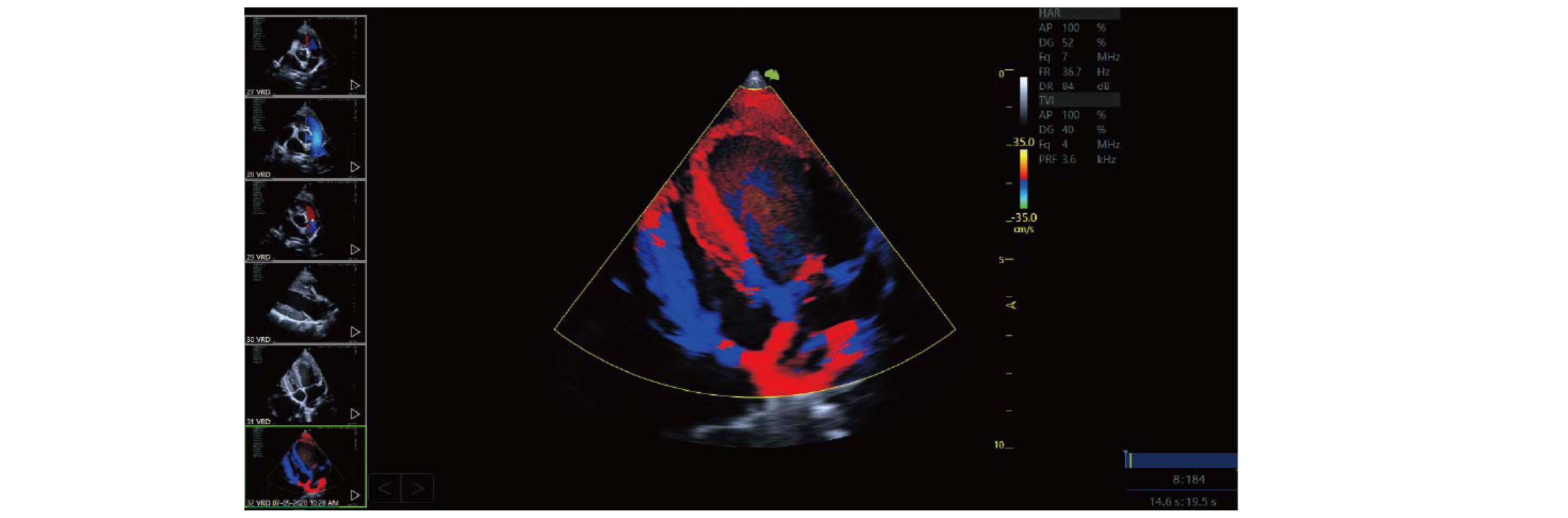
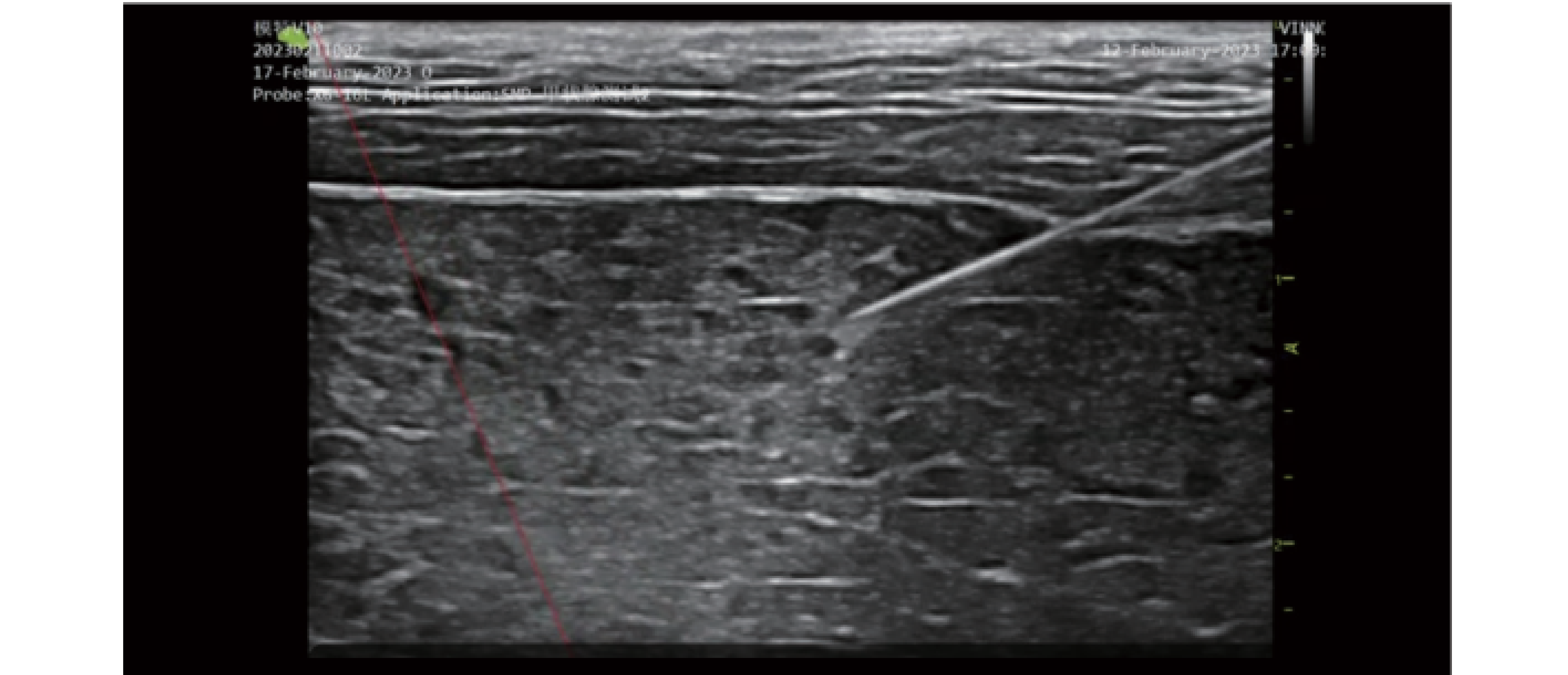
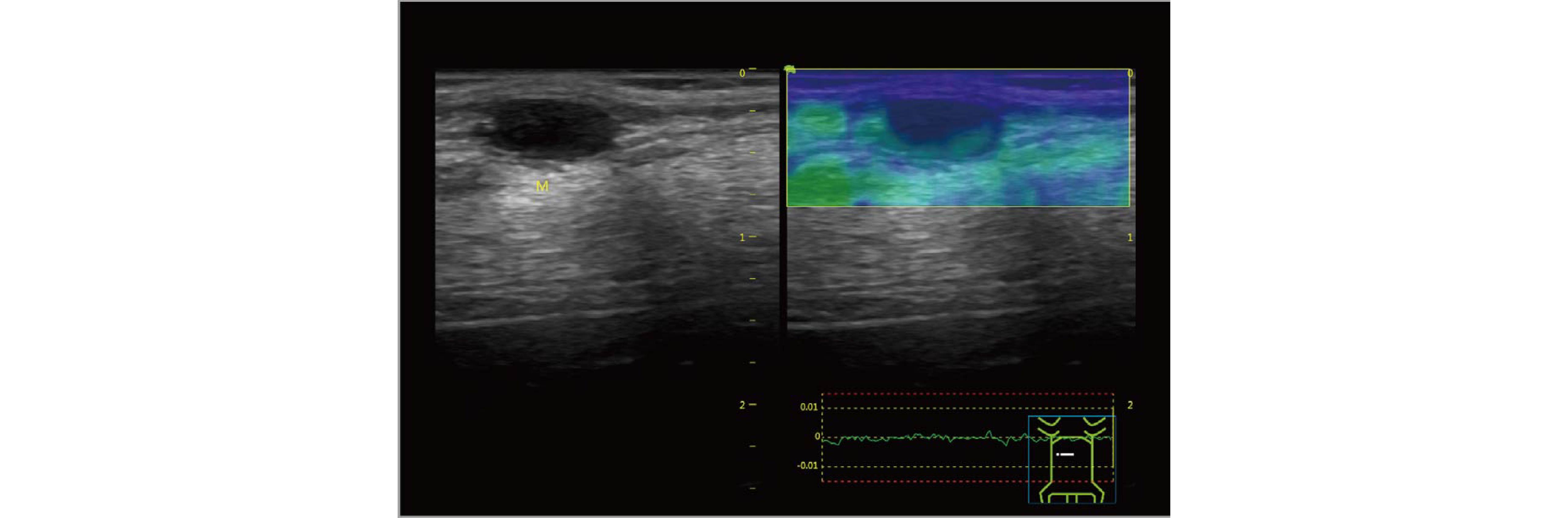
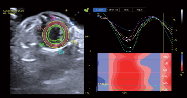
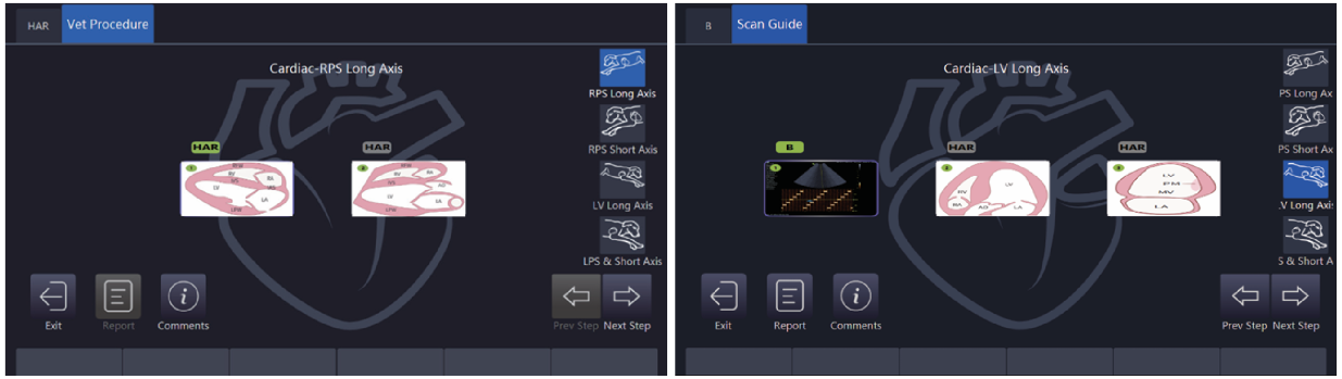
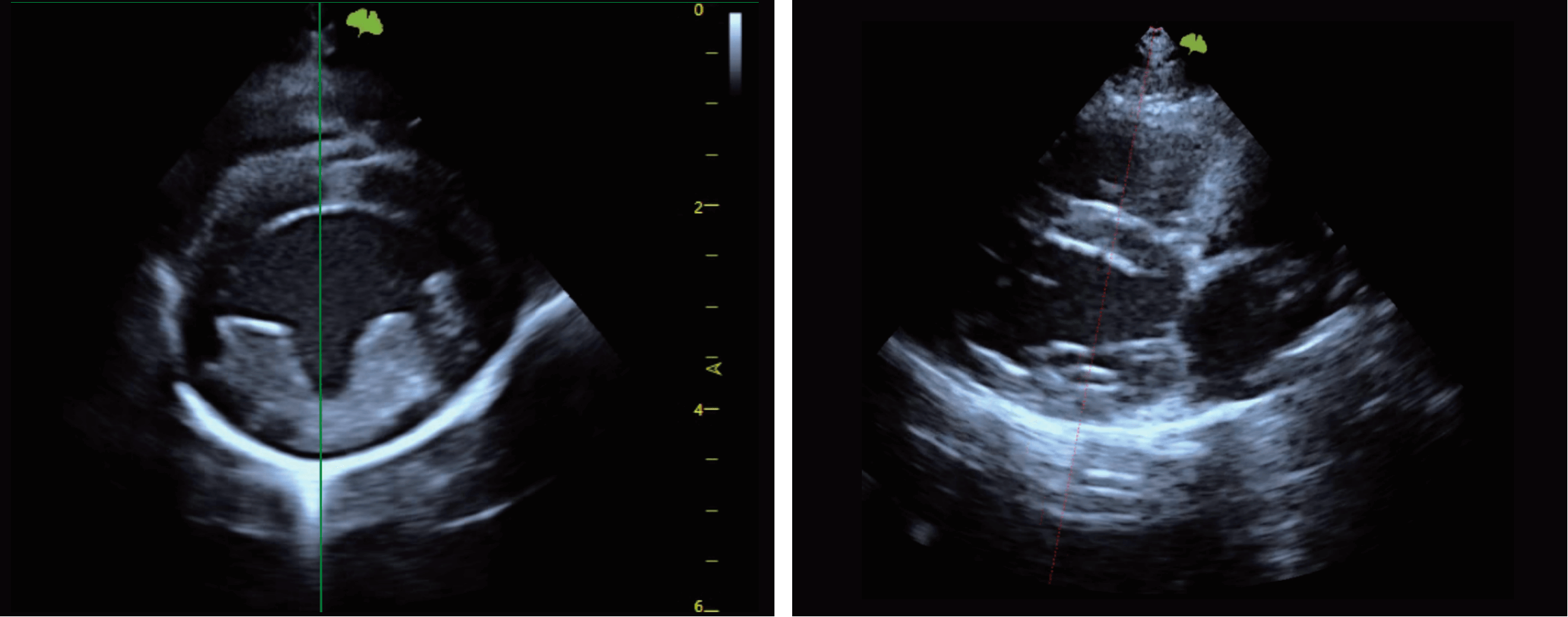
The VINNO D10 portable ultrasound system has been thoughtfully designed to overcome the challenges from everyday healthcare environments. With enhanced image quality based on VLucid+ platform, well designed workflow and smart automation tools, VINNO D10 provides the best-in-class image quality.

Advanced Features

Velp

The Al-powered Velp delivers real-time recognition of five standard cardiac planes with color-coded guidance for precise scanning. Automated measurements streamline diag-nostics, enhancing accuracy and empowering veterinarians to make faster, more informed clinical decisions.

VINNO Cardiac Quantification (vCQ)

The vCQ decision-making support alerts veterinarians to potential cardiac measurement issues by flagging values that fall outside predefined ranges. It enhances diagnostic accuracy, allowing veterinarians to quickly identify and address abnormalities.

Scan Guide & Tutorial

Provides standardized guidance for cardiac scanning with automated image saving, along with anatomical illustrations, transducer positioning videos, standard ultrasound videos, and scanning tips-supporting both abdominal and cardiac examinations.
Premium Cardiovascular solutions
Strain imaging
Quantify diastolic and systolic left ventricular function, evaluate cardiac strain
Velocity Vector Imaging (VVI)
Delivers more robust motion analysis than strain, enabling earlier and more precise detection of dysfunction and dyssynchrony
Multi Doppler
An unique technology that displays two doppler waveforms from separate sample gates during the same cardiac cycle. The optional modes include PW/PW, PW/TD, TD/TD
Advanced Features
Contrast Bubble Imaging
Optimized contrast imaging mode designed to enhance microcirculation visualization. With high sensitivity and real-time performance, CBI improves the depiction of fine vascular structures during contrast-enhanced examinations.
Needle Enhancement
Improves needle visibility during ultrasound-guided procedures, supporting clearer visualization of needle position in real time.
Elastic imaging
Provides visual assessment of tissue stiffness, supporting differentiation of tissue characteristics during ultrasound examinations.
Cutting-edge AI technology
VVI
Added VVI function, Users can set the display density before doing spot tracking.
Scan Guide
After entering the scanning guidance interface, the system has a built-in cardiac scanning guidance process. The flow can help junior physicians to scan slices according to the appro priate process. When the user clicks next, the VRD image data of the current scan will be automatically saved.
Velp
The user first selects a cutaway to scan, and then the AI algorithm examines each frame of the image. If the correct cutaway is not scanned, a red dashed line is displayed. When the user scans the correct cutaway, a green dashed line is displayed on the image
Ergonomics
01
Built-in camera & microphone for remote solution
02
15.6" monitor
03
8" touch screen
04
External multi-functional holder
05
2 USB + 1 Type-C port
06
Built-in ECG port
07
"Touch-and-view" external battery status display (when powered off)
08
2 USB + 1 Type-C port
Image Gallery
Contact us for any questions
Get in touch