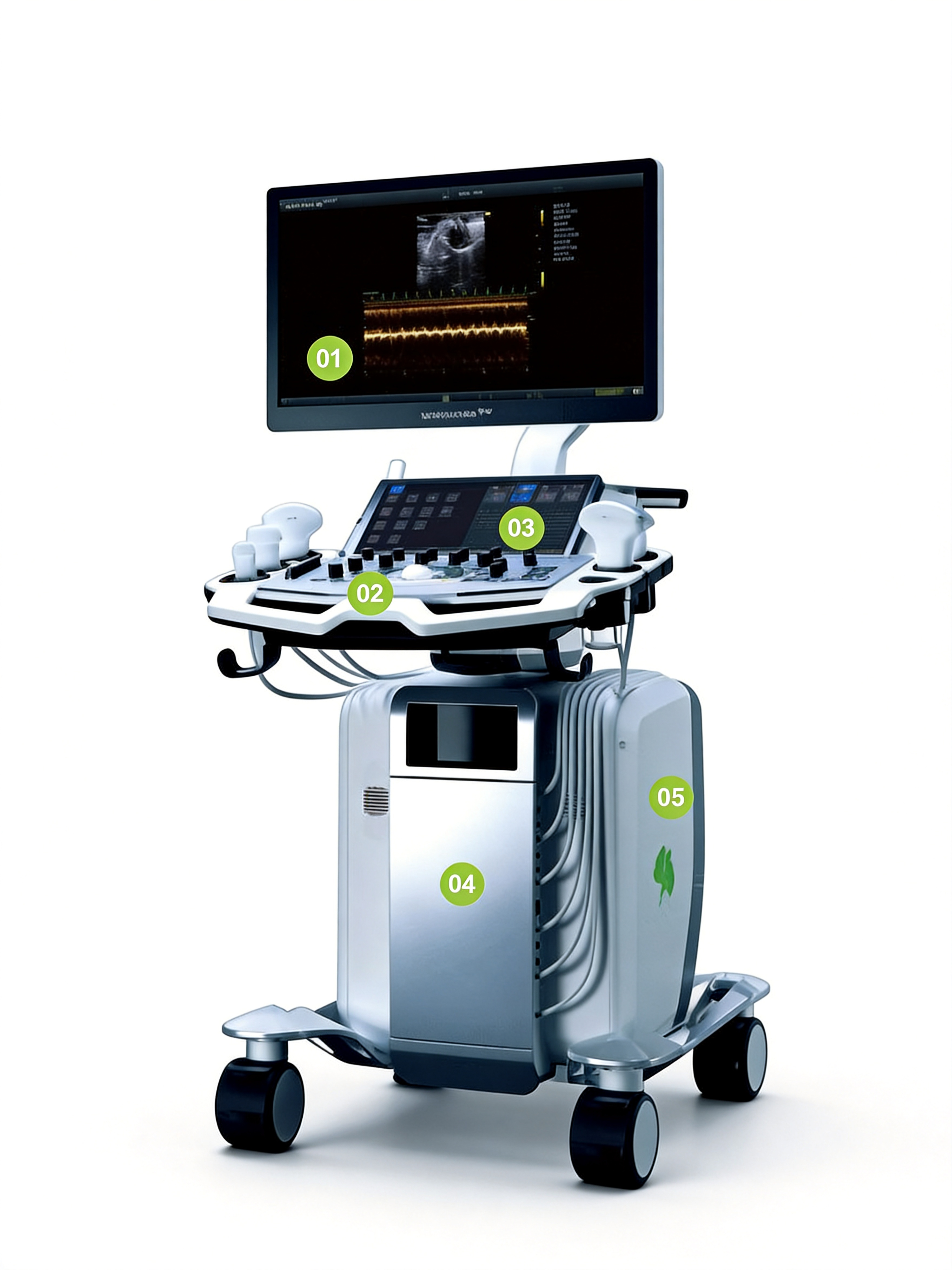
ULTIMUS 9LAB
Your Laboratory Partner
Research-Optimized Imaging Capabilities

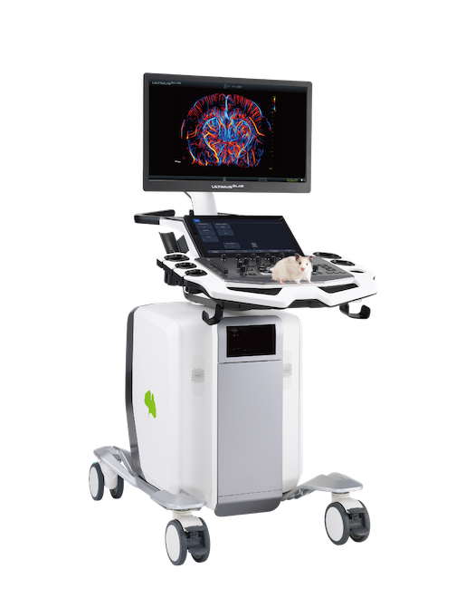
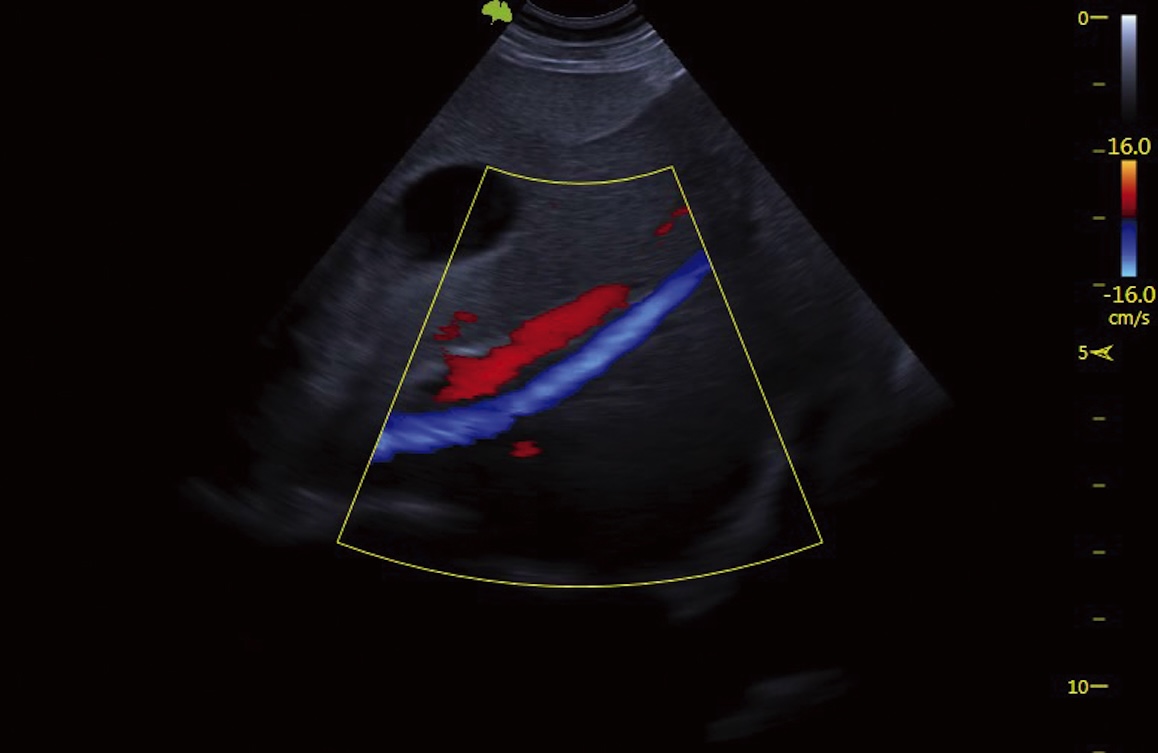
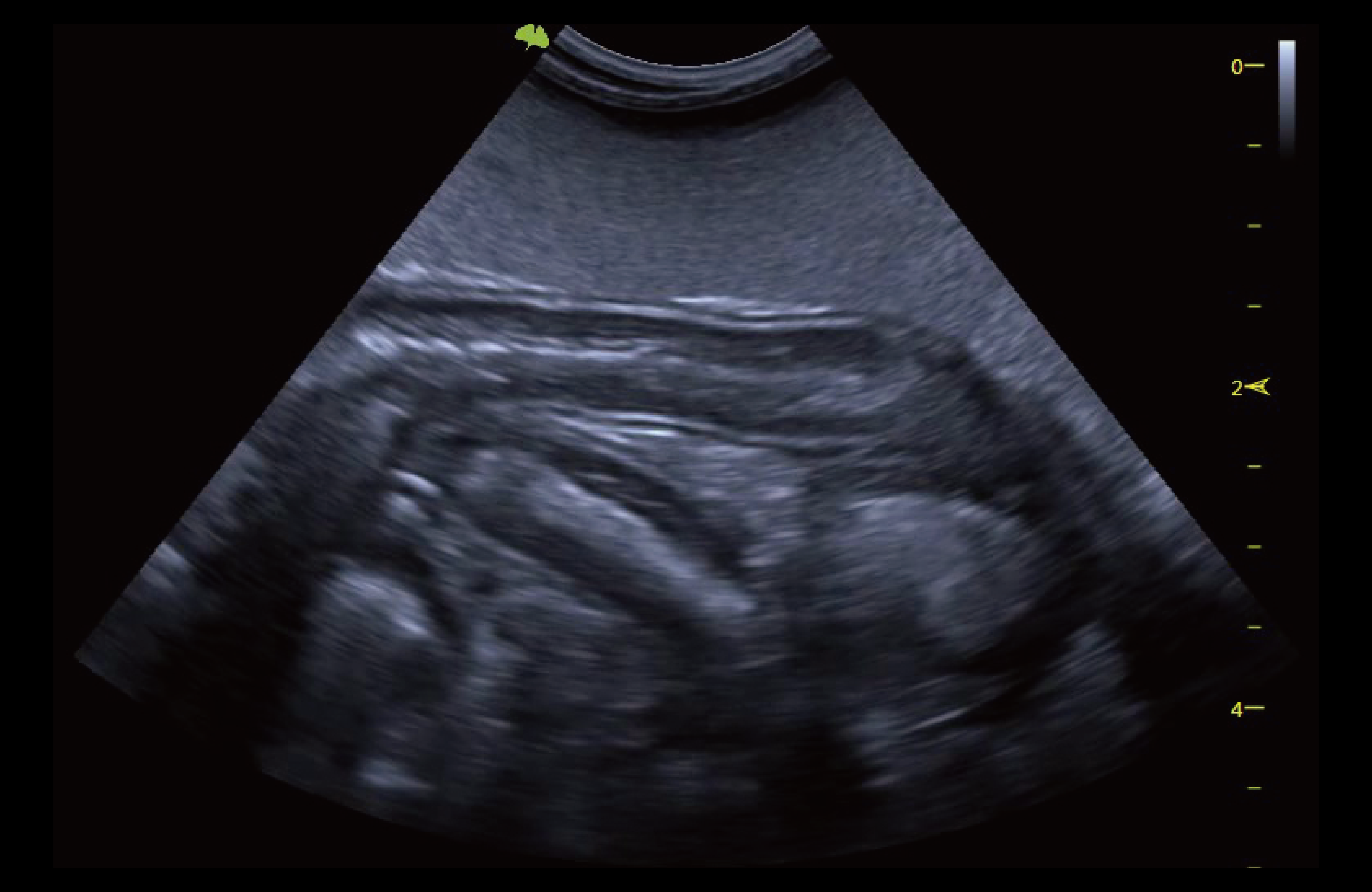
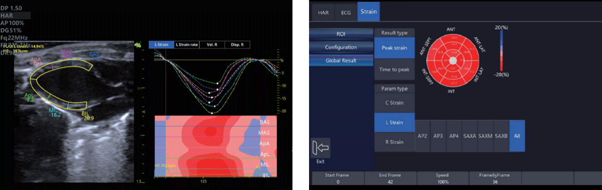
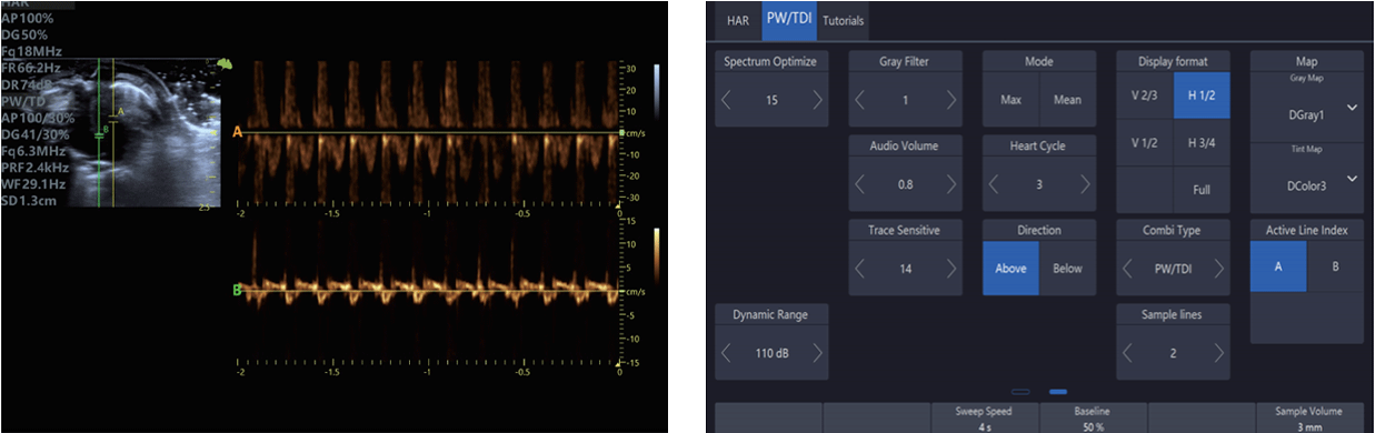
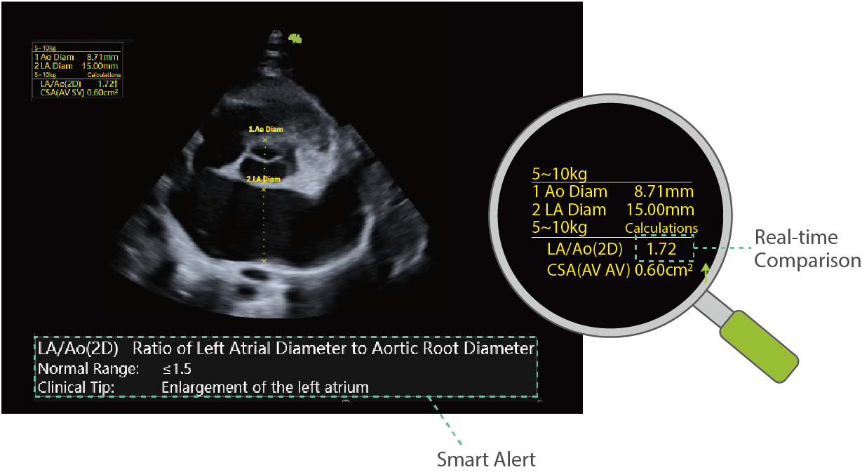
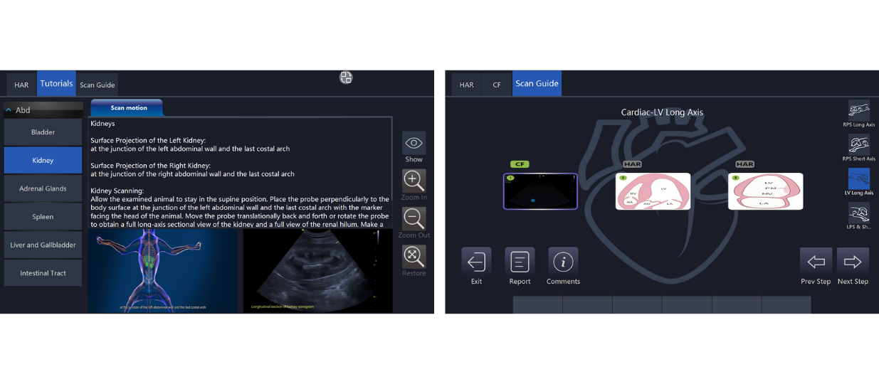
Built on the revolutionary MUSE platform with imaging frequencies up to 40 MHz, Ultimus 9LAB is purpose-built for laboratory and preclinical research. Lab-tailored presets, dedicated research ultrasound modes, and comprehensive quantification tools support advanced imaging across a wide range of animal models.


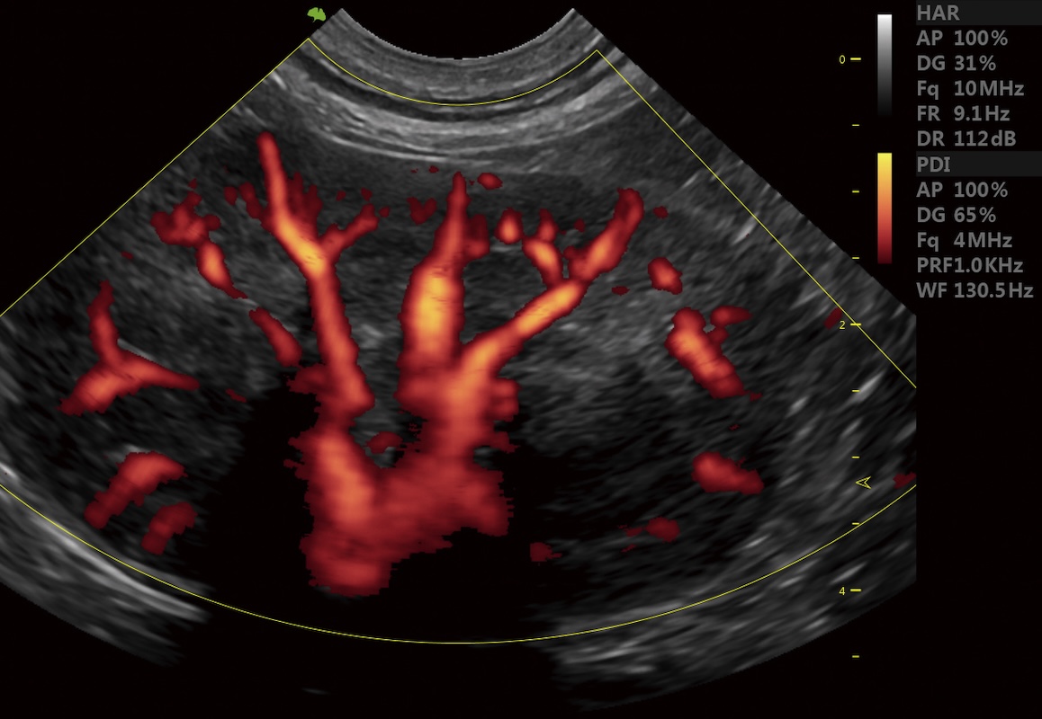
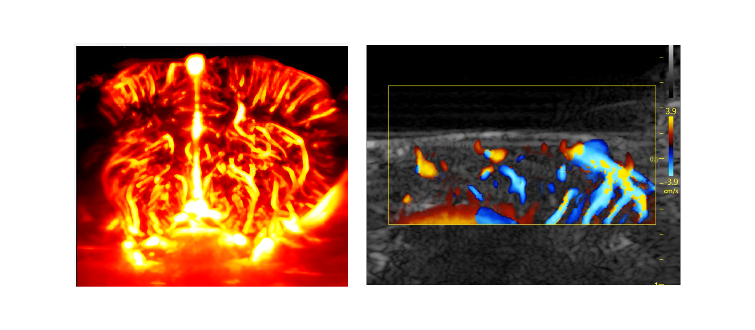
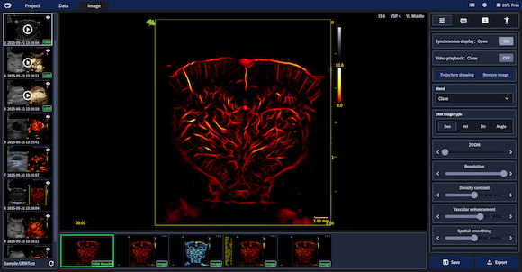
Integrated Ultra-Resolution Microscopy (URM) delivers micron-scale microvascular visualization and quantitative analysis, making Ultimus 9LAB an ideal engine for cutting-edge research workflows.

    - Lab-purpose: Lab-specific presets and quantification

    - URM: Ultra-Resolution Microscopy

    - 40MHz high-frequency


Ultra-Resolution Microscopy (URM)
With 10x micron-scale resolution, URM visualizes microcirculation in exceptional detail and delivers 10+ microvascular metrics-such as vessel density, velocity, flow direction, complexity, and tortuosity to support earlier evaluation and advance laboratory research. - Brain Science Research - Tumor Research - Microcirculation Research - URM Analysis (Tumor heterogeneity analysis): Automatic division of intratumoral and peritumoral region definition, to evaluate vascular morphology and hemodynamic parameters in different regions of the tumor
Super Micro Flow (SMF)
Detects ultra-fine blood flow and slow velocity in small vessels without using contrast agents, overcoming limitations of traditional flow modes.
Advanced Features for Lab Scenario

VFlash: Low-intensity ultrasound cavitation therapy

Under ultrasound, microbubbles undergo contraction and expan-sion, supporting applications in tumor chemo- and radiotherapy, myocardial perfusion enhance-ment, ultrasonic thrombolysis, and targeted drug delivery.

Contrast Bubble Imaging (CBI)

Contrast imaging optimized for microcirculation. An advanced, highly sensitive contrast bubble imaging which enhances real-time vessels visualization significantly based on ultra-fast frame rate.

VShear

Powered by Muse Platform, VShear allows more sensitive tissue detection by capturing every shear wave motion detail at ultrafast frame rates, which delivers great stability and accuracy during live scanning.

Flyinsono Lab

Flyinsono Lab is a preclinical research platform that seamlessly integrates with small-animal imaging systems for unified data transfer, storage, measurement, and case management. By shifting analysis to the PC, it frees researchers from long supervision and supports offline, batch, and automated processing-streamlining workflows and accelerating research. Key functions: URM offline analysis, cardiac quantification, automated processing, data export & reports, collaborative workflow, account isolation security
Premium Cardiovascular solutions
Total solution for cardiovascular health with high frequency imaging
Strain imaging
Quantify diastolic and systolic left ventricular function,Evaluate cardiac strain
Multi Doppler
A technology that displays two Doppler waveforms from separate sample gates during the same cardiac cycle.The optional modes include PW/PW,PW/TDI, TDI/TDI
Velocity Vector Imaging (VVI)
Delivers more robust motion analysis than strain, enabling earlier and more precise detection of dysfunction and dyssynchrony
Integrated Companion Animal Imaging
AI Solution in Cardiac Scanning
Velp
The Al-powered Velp delivers real-time recognition of five standard cardiac planes with color-coded guidance for precise scanning. Automated measurements streamline diag-nostics, enhancing accuracy and empowering veterinarians to make faster, more informed clinical decisions.
VINNO Cardiac Quantification (vCQ)
The vCQ decision-making support alerts veterinarians to potential cardiac measurement issues by flagging values that fall outside predefined ranges. It enhances diagnostic accuracy, allowing veterinarians to quickly identify and address abnormalities.
Scan Guide & Tutorial
Provides standardized guidance for cardiac scanning with automated image saving, along with anatomical illustrations, transducer positioning videos, standard ultrasound videos, and scanning tips-supporting both abdominal and cardiac examinations.
Special settings for lab mice mode
Supports multi-cycle myocardial strain imaging
Strain mode specially set up for laboratory mice
Ergonomic Design for Ultimate Convenience
01
Large-viewing Monitor
With 24” high-resolution LED monitor provides about 25% more lateral viewing information for a larger image display.
02
Space Capsule
The excellent heating dissipationability, anti-electromagnetic interference and noise isolation ensure the system deliver its best performance.
03
Highly-sensitive Touch screen
A high-sensitivity full-HD touch screen with 60 degree tiltable angle, custom-izable interface, to make viewing and workflow significantly easy.
04
Large-viewing Monitor
A high-sensitivity full-HD touch screen with 60 degree tiltable angle, custom-izable interface, to make viewing and workflow significantly easy.
Image Gallery
Bilobed Gallbaldder
Cardio PW
Cardio TVM
Zebra fish CF
Hepatic Vein
Intestinal Tract
Kidney PDI
Contact us for any questions
Get in touch